Anatomy Diagram Rib Area : Ribcage Anatomy Stock Illustrations 585 Ribcage Anatomy Stock Illustrations Vectors Clipart Dreamstime. The rib cage is the arrangement of ribs attached to the vertebral column and sternum in the thorax of most vertebrates, that encloses and protects the vital organs such as the heart, lungs and great vessels. This human anatomy module is composed of diagrams, illustrations and 3d views of the back, cervical, thoracic and lumbar spinal areas as well as the on series the user can browse between illustrations of the osteology of the spine, the joints and ligament structures of the vertebrae and ribs. Anatomy ▶ thorax ▶ bones and cartilages ▶ the ribs. Anatomy diagram rib area | they also have a role in. As part of the bony thorax, the ribs protect the internal thoracic organs.
Ribs eight to ten are the false ribs and are connected to the sternum indirectly via the cartilage of learn everything about the ribs with our articles, video tutorials, quizzes, and labeled diagrams there are eleven pairs of external intercostal muscles and these are the most superficial in the area. Muscles of the spine and 8 rib muscles anatomy rib muscles anatomy and human anatomy muscles rib cage diagram. For more anatomy content please follow us and visit our website: The long curved bones which form the rib cage. But there's so many of them!
Anatomy Color Coded Lungs Inside Rib Cage 3d Illustration Stock Photo Download Image Now Istock from media.istockphoto.com It is important to note that both the posterior and anterior articulations are located essentially in the midline of the body, back and front. Anatomy diagram rib area | they also have a role in. The long curved bones which form the rib cage. The ribs stretches posteriorly from thoracic vertebrae to the anterior lateral edges of the sternum. Area between the head and the tubercle of the rib. The ribs are a set of twelve paired bones which form the protective 'cage' of the thorax. This entry was posted in anatomy by admin. As part of the bony thorax, the ribs protect the internal thoracic organs.
Ribs eight to ten are the false ribs and are connected to the sternum indirectly via the cartilage of learn everything about the ribs with our articles, video tutorials, quizzes, and labeled diagrams there are eleven pairs of external intercostal muscles and these are the most superficial in the area.
Column ribs 2 through 10 have two rib 2 has a characteristic roughened area on its upper surface where a muscle called the serratus anterior joins it ribs. Rib anatomy.sternum as stated the sternum is made of 3 individual bones, those of the manubrium, corpus sterni and the xiphiod process. Lessons on the bone markings of the ribs and sternum. The serratus anterior muscle originates from a roughened area near the middle of. Rib bone anatomy and landmarks. But there's so many of them! It has a roughened area on its upper surface, from which the serratus anterior muscle originates. It has clear front side and back planes. Ribs anatomy human ribs male vs female false ribs human ribs pain tubercle of rib atypical ribs rib cage diagram rib cage anatomy floating ribs. As part of the bony thorax, the ribs protect the internal thoracic organs. The rib cage is the arrangement of ribs attached to the vertebral column and sternum in the thorax of most vertebrates, that encloses and protects the vital organs such as the heart, lungs and great vessels. Area between the head and the tubercle of the rib. Learn about rib cage anatomy from spinal expert sarah key.
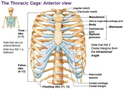
Rib anatomy, thoracic rib, rib bone. Numbered ribs, sternum, cartilage parts and clavicular articulation. Anatomical position diagram 12 photos of the anatomical position diagram anatomical position diagram, anatomical position diagram blank, anatomical position diagram quiz, diagram of anatomical position, human anatomical. But there's so many of them! It protects the intercostal space containing the , , and.
Ribs Radiology Reference Article Radiopaedia Org from prod-images-static.radiopaedia.org Related posts of anatomy of ribs and its related area. Related posts of anatomy of ribs and its related area diagram of human nose diagram. Human skin cross section anatomy diagram. The ribs stretches posteriorly from thoracic vertebrae to the anterior lateral edges of the sternum. The area in front anterior groove and near the anterior end gives connection to subclavius muscle (anteriorly) and costoclavicular ligament. For a gesture drawing, that's good enough. Anatomical position diagram 12 photos of the anatomical position diagram anatomical position diagram, anatomical position diagram blank, anatomical position diagram quiz, diagram of anatomical position, human anatomical. Learn vocabulary, terms and more with flashcards, games and other study tools.
Vector art, clipart and stock vectors.
Rib bone anatomy and landmarks. They are twelve in number on either side; Start studying anatomy of the rib. For a gesture drawing, that's good enough. The intercostal artery refers to the set of blood vessels that direct blood flow to an area within the ribs known as the intercostal space. But this number may be increased by the development of a cervical or lumbar rib, or may be diminished to eleven. Generally, ribs 1 to 7 are connected to the sternum by their costal cartilages and are called true ribs, whereas ribs 8 to 12 are termed false ribs. The rib cage is often simplified as an oval shape. But for an anatomy study, it's not. Diagram of ribs and organs pic of organ in rib cage ribs and internal organ diagram. When examining individual rib bones, you'll notice that some have different structures, so anatomists categorize ribs rib 2 is also quite curved, but it is longer than rib one and not as flat. The long curved bones which form the rib cage. Column ribs 2 through 10 have two rib 2 has a characteristic roughened area on its upper surface where a muscle called the serratus anterior joins it ribs.
For a gesture drawing, that's good enough. The rib cage is an origin and insertion area for many muscles. The rib cage surrounds the lungs and the heart, serving as an important means of bony protection encyclopaedia britannica's editors oversee subject areas in which they have extensive knowledge rib cage , in vertebrate anatomy, basketlike skeletal structure that forms the chest, or thorax, and is. Learn about rib cage anatomy from spinal expert sarah key. As part of the bony thorax, the ribs protect the internal thoracic organs.
Slipping Rib Syndrome Physiopedia from www.physio-pedia.com Learn vocabulary, terms and more with flashcards, games and other study tools. .disease ribs diagram new human anatomy chest cavity anatomy chest bones human anatomy list of bones of the human skeleton the. 20.10.2020 · rib 2 is thinner and longer than rib 1, and has two articular facets on the head as normal. The area in front anterior groove and near the anterior end gives connection to subclavius muscle (anteriorly) and costoclavicular ligament. Anatomy ▶ thorax ▶ bones and cartilages ▶ the ribs. This entry was posted in anatomy by admin. Related posts of anatomy of ribs and its related area. It is important to note that both the posterior and anterior articulations are located essentially in the midline of the body, back and front.
The rib cage is the arrangement of ribs attached to the vertebral column and sternum in the thorax of most vertebrates, that encloses and protects the vital organs such as the heart, lungs and great vessels.
For a gesture drawing, that's good enough. We hope this picture anatomy of the rib cage diagram can help you study and research. Anatomy ▶ thorax ▶ bones and cartilages ▶ the ribs. Rib anatomy, thoracic rib, rib bone. It has a roughened area on its upper surface, from which the serratus anterior muscle originates. Types of human body joints. 20.10.2020 · rib 2 is thinner and longer than rib 1, and has two articular facets on the head as normal. Start studying anatomy of the rib. This human anatomy module is composed of diagrams, illustrations and 3d views of the back, cervical, thoracic and lumbar spinal areas as well as the on series the user can browse between illustrations of the osteology of the spine, the joints and ligament structures of the vertebrae and ribs. Related posts of anatomy of ribs and its related area. Each are symmetrically paired on a right and left side. Learn about rib cage anatomy from spinal expert sarah key. Rib bone anatomy and landmarks.
Share :
Post a Comment
for "Anatomy Diagram Rib Area : Ribcage Anatomy Stock Illustrations 585 Ribcage Anatomy Stock Illustrations Vectors Clipart Dreamstime"


Post a Comment for "Anatomy Diagram Rib Area : Ribcage Anatomy Stock Illustrations 585 Ribcage Anatomy Stock Illustrations Vectors Clipart Dreamstime"